ZEISS - Primo Vert
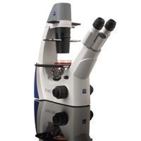
Quick Assessment and Inspection of Living Cells
Utilize Primo Vert in routine laboratories for life cell inspection. Also in cutting edge research laboratories Primo Vert is a reliable routine microscope for a quick and efficient check of living cells. Typical applications include e. g. cancer and HIV research, human, animal and plant genetics and cell biology in general.
Active Questions & AnswersAsk a Question
Flashing power lights
Recent Questions & Answers
Updated byIlluminators
Need Equipment Support?
Documents & ManualsView All Documents
Features of Primo Vert
- Automatic light turn-off
- Modular illumination: HAL or LEDs
- Universal phase slider for all objectives
- Robust and durable
- Increased working distance, e. g. for roller-bottles
- Carrying handle on the back
- Objective-indicators for fast identification of magnification
General Specifications
| Microscope Type | Inverted |
Additional Specifications
Light Sources
Halogen lamp: HAL 6 V, 30 W
Adjustability of light source: continuous, from 1.5 to 6 V DC
Color temperature at 6 V: 2800 K
Luminous flux: 765 lm
Average life: 100 h
Luminous area: 1.5 x 1.5 mm
LED illumination
White light LED, peak wavelength 450 nm,
LED risk group 2 according to IEC 62471
Constant, brightness-independent color temperature: 7480 K
Homogeneous field illumination: 20 mm diameter
Suitable for objectives with magnifications of: 4x to 40x
Analogous brightness adjustment from: approx. 15 to 100 %
Optical/Mechanical Data
Stand with stage focusing drives
With coarse focusing drive: 45 mm/rev.
With fine focusing drive: 0.5 mm/rev.
Total stage lift: 15 mm
Optics
Objective change: manual via quadruple objective nosepiece
Objectives: infinity-corrected objective range with W 0.8 mounting thread
Eyepieces: 30 mm tube diameter
with field-of-view number 20: WF 10x/20 Br. foc.
Specimen Stage
Specimen stage: fixed
Dimensions (width x depth): 200 x 239 mm
Specimen guide: right side
Verniers with numerical and alphabetic scale: X direction: numerical scale, readable from right to left
Coaxial drive: Y direction: alphabetic scale, readable in the mirror right side
Condensers
LD condenser 0.3: for Vobj 4x to 40x, a = 72 mm
LD condenser 0.4: for Vobj 4x to 40x, a = 55 mm
Versions of Stands
Binocular tube 45°/20
Maximum field-of-view number: 20
Interpupillary distance: adjustable from 48 to 75 mm
Tube angle: 45°
Viewing height: 360 to 397 mm
Viewing port: tube factor 1x
Trinocular (photo)tube 45°/20
Maximum field-of-view number: 20
Interpupillary distance: adjustable from 48 to 75 mm
Tube angle: 45°
Viewing height: 360 to 397 mm
Viewing port: tube factor 1x
Photo/video port: tube factor 1x, 60 mm mount
Fixed beam splitting: 50 % vis / 50 % doc
Primo Vert Monitor
Camera: 5 Megapixel CMOS
Monitor size: 8,4”
Display: 800x600 Pixel
Storage medium: Secure Digital (SD)-card
Outputs/ports: USB
Camera driver for: AxioVision LE with special
Microscopy software: configuration too