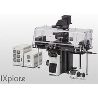
Olympus - IXplore Live
No Documents
Precise Live Cell Imaging
Active Questions & AnswersAsk a Question
There are no current Discussions
Need Equipment Support?
Documents & Manuals
There are no Documents or Manuals available.
Features of IXplore Live
Meeting the Needs of Live Samples Live cells require careful maintenance of their environment to grow and thrive, and Olympus offers a variety of microscope-based incubation systems to meet evolving research needs. Box-type incubation systems* enable time-lapse observations over a period of several days by enclosing a portion of the microscope within the incubator. Shorter time-lapse experiments can be completed with stage-top microscope CO2 incubation systems* that can be fitted to the stage and easily removed when not in use.
Both systems are precisely controlled to maintain a constant environment surrounding the dish or well plates, controlling temperature, humidity, and CO2 concentration. This maintains cell activity to significantly improve the reliability of time-lapse observation and provide better data.
Constant Environmental Control In this SelectScience interview, Jutta Bulkescher, Microscopy Specialist at the Center for Protein Research/Danish Stem Cell Center, University of Copenhagen, describes the wide range of research conducted at her facility and explains how the Olympus cellVivo incubation system enables her to reliably perform stem cell analysis, while maintaining cells under strict physiological conditions.
Hardware Stability The frame architecture and focus drive design of the IXplore system offer enhanced rigidity that reduces the impact of vibration and temperature on the microscope. This helps maintain desired positions along the X, Y, and Z axes to facilitate reliable time-lapse and multipoint imaging. When using the IXplore Live system combined with the Olympus ultrasonic stage (IX3-SSU) and TruFocus system, you can capture high-precision, multipoint time-lapse images that are aligned and in focus.
Live Cell Imaging Olympus silicone oil immersion objectives provide clearer images of living specimens during time-lapse experiments. The refractive index of silicone oil (ne?1.40) is close to that of living tissue (ne?1.38), so these objectives help reduce the spherical aberration caused by refractive index mismatch, enabling high-resolution observation deep inside living tissue. Silicone oil does not dry out or harden, so there is no need to add more oil, making it ideal for extended time-lapse observations.
Microsecond Accuracy Device Control Fast filter wheels, shutters, individual LED light source control, and real-time controllers (U-RTC) help reduce photobleaching and phototoxicity, resulting in healthier cells and more robust data.
Monitor Cell Migration and Growth Analyze the movement and division of live cells in time-lapse or z-stack image sets with cellSens Object Tracking and Count and Measure solutions. Use the Confluency Checker tools to measure confluency on phase contrast images in addition to fluorescence.
Rapid Deconvolution Olympus cellSens Dimension software includes live 2D deblurring for preview and acquisition to enable better focusing on thick specimens. More advanced TruSight deconvolution is available to reassign out-of-focus light through the CI deconvolution solution. TruSight uses a constrained iterative deconvolution algorithm to produce improved resolution, contrast, and dynamic range with high speed through GPU processing.
Large Field of View The large field of view Olympus optics, including mirror units and fly-eye lens systems, provide uniformly illuminated fluorescence images and enable the use of sCMOS cameras with large sensors.
Ease of Use The Graphical Experimental Manager (GEM) of cellSens Dimension software offers fully automated multidimensional observation (X, Y, Z, T, wavelength, and positions) and eases experiment setup.
General Specifications
There are no General Specifications available.