HORIBA - DeltaMyc
Time-resolved fluorescence microscopy is the ultimate tool to study dynamic events in cellular structures and nanomaterials.
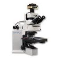
The DeltaMyc features time-correlated single photon counting (TCSPC) for sensitive and rapid acquisition of luminescence lifetimes from 100 ps to seconds. It is composed of a compact optical module added to an Olympus BX 53 upright microscope or IX73 inverted microscope. Its mapping capabilities include an automated X,Y fast scanning stage, which combined with its confocal ability, can generate fluorescence lifetime mapping with micron level spatial resolution.
The DeltaMyc is a flexible research tool that combines a large range of picosecond pulsed laser diode sources spanning wavelengths from 370 to 980 nm and repetition rates from CW to 100 MHz, multiple filter configurations and various detector options to suit your specific measurement needs. An included high dynamic range, low noise, cooled camera and a high intensity fluorescence illuminator configured with the DeltaMyc enable widefield fluorescence imaging.
The DeltaMyc features fully-automated mapping controlled from the intuitive user interface of our DataStation software, while retaining the full flexibility and functionality of the microscope. Full reconvolution analysis can be performed to generate maps of the fit parameters such as lifetimes, relative amplitudes, average lifetime, and fluorescence intensity. For example, it is ideal an instrument study protein dynamics such as binding or disassociation using Fluorescence Resonance Energy Transfer (FRET).
Active Questions & AnswersAsk a Question
There are no current Discussions
Fluorescence Detector Service ProvidersView All (21)
Documents & ManualsView All Documents
Features of DeltaMyc
- Single photon counting (TCSPC) for lifteimes from 100 ps to seconds * Large selection of various laser diodes sources (370 – 980 nm), dichroic filters and pinholes to match any sample * New laser diode (DeltaDiode™) with high repetition rate lasers (up to 100MHZ), operated as CW or pulsed mode * Intuitive data acquisition and analysis software * FRET software routine for transfer efficiency and molecular distance * Cooled fluorescence camera for wide field fluorescence imaging
General Specifications
There are no General Specifications available.
Additional Specifications
Microscope
Based on upright Olympus BX53 microscope or IX73 inverted microscope.
Objectives
Plan achromat 10× and 60×, other magnifications available. 5-position turret.
Confocal emission pinhole
5 diameters, from 100 Μm to 1000 Μm, manually selected one at a time.
Camera
Fluorescence camera
1.4 Mpix cooled
Excitation sources
Direct-coupled pulsed laser sources
Wavelength range
Any DeltaDiode source available
Motorized stage
Resolution
1.0 Μm
Travel range
75 × 50 mm
Manual control
With joystick
Automatic control
Through DataStation software
TCSPC electronics
Single-photon counting detection
Resolution
26 ps/channel
Lifetime range
100 ps to 10 Μs, depending on sample
Detector
PPD fast PMT
Spectral range
250–650 nm / 300–850 nm
Transit spread time
200 ps
Dark counts
< 80 cps
Filters
5 manually selected positions of interchangeable cubes which can hold:
Excitation filters
Optional 10 nm BP filters or short-pass filters
Dichroic beam splitters
Optional wavelength or 50/50
Emission filters
Optional 40 nm BP filters or long-pass filters
ND filters
5 positions: 0, 0.3, 1, 2, and 3 OD
Manually selected
Software
Data acquisition
DataStation software
TCSPC mapping
High-speed scan
Data analysis
DAS6 software, inc. reconvolution feature
Operating system
Windows® 7
Dimensions
<25 cm added to top of microscope