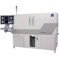
ZEISS - Xradia 520 Versa
Submicron Imaging with New Degrees of Freedom
ZEISS Xradia 520 Versa 3D X-ray microscope unlocks new degrees of flexibility for scientific discovery. Building on industry-best resolution and contrast, Xradia 520 Versa expands the boundaries of non-destructive imaging for breakthrough flexibility and discernment critical to your research. Innovative contrast and acquisition techniques free you to seek-and find-what you've never seen before to move beyond exploration and achieve discovery.
Xradia 520 Versa achieves spatial resolution of 0.7 µm and minimum achievable voxel of 70 nm.
The most comprehensive submicron X-ray imaging solution offering unprecedented flexibility
In addition to well-known advantages offered by the ZEISS Xradia Versa series of X-ray microscopes – highest resolution, best contrast, RaaD (resolution at a distance) and non-destructive submicron X-ray imaging – ZEISS Xradia 520 Versa advances lab-based X-ray imaging with breakthrough techniques and innovations:
Dual Scan Contrast Visualizer (DSCoVer)
DSCoVer provides flexible side-by-side tuning of two distinct tomographies at different imaging conditions or sample conditions. This enables compositional probing for features normally indistinguishable in a single scan, enabling you to seamlessly and easily collect the data required for dual energy analysis. Imaging a sample at two different X-ray spectra, or in two different states, aligning the resulting datasets and then combining them assures you will achieve optimum contrast for the material of interest and allow you to establish repeatable research parameters.
DSCoVer takes advantage of how X-rays interact with matter based on effective atomic number and density. This provides you with a unique capability for distinguishing, for example, mineralogical differences within rocks as well as among difficult-to-discern materials such as silicon and aluminum.
High-Aspect Ratio Tomography (HART)
The innovative High Aspect Ratio Tomography (HART) mode on Xradia 520 Versa provides you with higher throughput imaging for flat samples such as those found with semiconductor packages and boards. HART enables you to space variable projections so that you collect fewer projections along the broad side of a flat sample and more along the thin side. A significant amount of 3D data is provided by these closely-spaced long views versus less densely-spaced short views.
You can also tune HART to emphasize higher throughput or better image quality, thereby potentially accelerating image acquisition speed by 2X.
Automated Filter Changer (AFC)
Now it’s even easier to image challenging samples. Source filters used to tune the X-ray energy spectrum are an important complement to DSCoVer and in situ applications. The AFC houses a standard range of 12 filters along with space for a dozen additional custom filters for new applications. Filter selection is easily programmed and recorded within imaging recipes so filters may be changed without disrupting the work flow.
Wide Field Mode & Vertical Stitching
Wide Field Mode provides you with either an extended lateral field of view for imaging large sample types or higher resolution using the standard field of view, both in a single tomography. For larger samples, the lateral field of view is approximately twice as wide as the standard mode providing 3D volume more than three times larger. Using standard field of view, WFM provides nearly twice the number of voxels. Combining WFM with the existing Vertical Stitching feature enables you to image large samples that are both wider and taller than the standard field of view.
Optional Diffraction Contrast Tomography (LabDCT)
Building on the industry-best resolution and contrast of ZEISS Xradia Versa X-ray microscopes (XRM), optional ZEISS LabDCT™ advanced imaging module provides you with direct visualization of 3D crystallographic grain orientation. This powerful combination of diffraction capability with unique reconstruction software for the Xradia 520 Versa meets the growing need for metallurgists and materials scientists to obtain such information to accompany your data sets. And now, for the first time ever, LabDCT brings non-destructive diffraction contrast tomography (DCT) directly to your laboratory.
Optional Flat Panel Extension (FPX)
ZEISS FPX™ flat panel extension enables unmatched versatility in a single X-ray system. Image significantly larger samples (beyond 5” diameter) and greater volumes (10X increase) with high throughput (better by 2-5X). Get to results faster by performing rapid macroscopic scouting scans and then zooming in to regions of interest at high resolution. With FPX, simplify and expand your multiscale imaging, characterization and modeling workflows all on a single platform.
Optional Autoloader
Maximize your instrument’s utilization by minimizing user intervention with the optional ZEISS Autoloader, available for all instruments in the ZEISS Xradia Versa series of 3D X-ray microscopes. Reduce the frequency of user interaction and increase productivity by enabling multiple jobs to run. Load up to 14 samples, queue, and allow to run all day, or off-shift. The software provides you with the flexibility to re-order, cancel and stop the queue to insert a high priority sample at any time. An e-mail notification feature in the ZEISS Scout-and-Scan user interface provides timely updates on queue progress. Autoloader also enables a workflow solution for high volume repetitive scanning of like samples.
Optional In Situ interface kit
X-ray imaging is uniquely suited to image materials under variable environments with controlled conditions including over time (4D) to non-destructively characterize and quantify the evolution of 3D microstructures. With its unique architecture, the Xradia Versa has emerged as the industry's premier solution supporting the use of the widest variety of in situ rigs, from high pressure flow cells to tension, compression and thermal stages. The optional In Situ Interface Kit delivers stable and robust management of often complex in situ electrical and plumbing facilities and ensures Xradia performance is maintained, along with recipe-based software capability that simplifies operation from within the Xradia Versa user interface. The In Situ Interface Kit is available on all Xradia Versa systems.
Active Questions & AnswersAsk a Question
There are no current Discussions
Microscope Cameras and Imaging Service ProvidersView All (19)
Documents & Manuals
There are no Documents or Manuals available.
Features of Xradia 520 Versa
The unique Xradia 520 Versa architecture delivers unsurpassed flexibility:
Unprecedented resolution in non-destructive 3D X-ray imaging
True spatial resolution <700 nm * Below 70 nm minimum achievable voxel * Two-stage magnification that provides Resolution at a Distance (RaaD), delivering large, flexible working distances while maintaining submicron resolution * Non-destructive interior tomography uniquely enabled by Scout-and-Zoom, now for samples 10x greater in volume with 2-5x greater throughput with the optional Versa FPX flat panel extension.
Advanced contrast capabilities for imaging challenging samples * Enhanced absorption contrast detectors maximize collection of contrast-forming low energy X-ray photons that are critical to imaging numerous material types * Tunable propagation phase contrast to visualize low Z materials and biological samples that tend to have limited absorption contrast * Maximum discernibility with dual energy probing of features normally indistinguishable within a single scan
The premier in situ and 4D solution * Nondestructive X-ray microscopes uniquely characterize the microstructure of materials in simulated conditions—in situ—as well as the evolution of properties over time (4D) * Supporting a wide variety of in situ rigs for submicron imaging of samples up to inches in size within environmental chambers and under varying conditions * RaaD enables Xradia Versa to maintain high resolution as the space between the X-ray source and sample grows whereas the resolution of conventional micro-CT architecture degrades when samples are placed within spacious in situ chambers * With Versa FPX, acquire images at high throughput in a large field of view, and then nondestructively sub-sample your region of interest with RaaD, all in one system
Further advantages * Wide Field Mode (WFM) nearly 2X as wide as the standard mode for larger field of view at resolution. Resulting 3D volumes are more than 3X larger. * Super simple control system for efficient workflows, ideal for a central lab-type setting where your users may have a wide variety of experience levels. * System stability: vibrational isolation, thermal stabilization, low noise detector. * Powerful dual GPU workstation that accelerates image reconstruction time by up to 40%. * Optional Versa In Situ Kit organizes the facilities that support environmental chambers (such as wiring and plumbing) to enable maximum imaging performance and ease set-up. * Autoloader option enables you to program and run up to 14 samples at a time to maximize productivity, automate workflows for high volume scanning.
General Specifications
There are no General Specifications available.

