Olympus - VS120
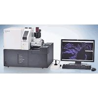
Virtual Slide Microscopy
The VS120 is not for clinical diagnostic use.
Active Questions & AnswersAsk a Question
There are no current Discussions
Need Equipment Support?
Documents & ManualsView All Documents
Features of VS120
Ideal for Research Facilities and Digital Learning
State-of-the-art Research Tool for Brightfield and Fluorescence * Advanced Medical Education and Collaboration * Remote Conferencing and Consultation
High-detail, High-precision, Rapid Scanning * Always in Focus with the VS120 Focus Map * Wide Range of Objectives from 2x to 100x * Virtual-Z with 3D Virtual Slide Production * Accurate Image Stitching, Regardless of Specimen Quality * High-resolution, High-sensitivity Virtual Fluorescent Slides * Automation Enhances Laboratory Efficiency
Intuitive slide scanning workflow and data review * Full Slide Scanning in Three Clicks of the Mouse * Instantly Get to the Desired Scanning Area * View Full and Magnified Images on the Same Screen * Innovative Synchronizing Enables Comparative Viewing of Different Stains * Save Voice Annotation Data * Capture Clear Images with All-in-Focus Imaging for Fluorescence
Powerful data management with NetImage Server SQL (optional) * Fast Data Access * SQL-based Net Image Server * Attach Metadata to Virtual Slides * Batch Management of Digital Content
Remote Access with Virtual Slide Viewers * OlyVIA Desktop * OlyVIA Mobile * Web Access
Darkfield Imaging * High-resolution Unstained and Fluorescent Images
General Specifications
| Microscope Type | Digital |
Additional Specifications
Intended Specimen
Observable Specimen: Glass slide with cover glass
Size of Glass Slide: Width: 25 mm - 26 mm, length: 75 mm - 76 mm, thickness: 0.8 mm - 1.4 mm
Size of Cover Glass: Thickness: 0.12 mm - 0.17 mm
Microscope Frame
Illuminator: Built-in Koehler illumination for transmitted light
Objectives: 2x,10x, 20x, 40x, 60x*1 and 100x*1 oil immersion with a motorized revolving nosepiece (* option only for VS120-S6-W)
Motorized Stage: Motorized XY stage with automatic control
Focusing: Motorized automatic control
Fluorescence Observation (Option): Motorized fluorescence illuminator, Motorized filter wheel, Fluorescence light source (Mercury lamp: U-HGLGPS or LED: Lumencor SOLA*2), Digital monochrome camera (Olympus XM10 or Hamamatsu ORCA Flash4.0 V2)
Digital Camera
CCD Camera: 2/3” CCD camera, 3.45 Μm x 3.45 Μm pixel size, high sensitivity, high resolution
Image Correction: Shading correction, auto white balance