ZEISS - Atlas 5
Manufactured by ZEISS
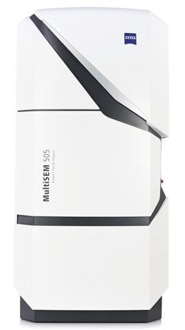
Atlas 5 makes your life easier: create comprehensive multi-scale, multi-modal images with a sample-centric correlative...
Atlas 5 makes your life easier: create comprehensive multi-scale, multi-modal images with a sample-centric correlative environment.
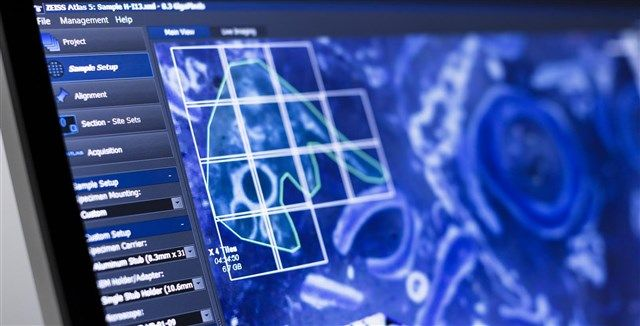
Atlas 5 is your powerful hardware and software package that extends the capacity of your ZEISS scanning electron microscopes (SEM) and ZEISS focused ion beam SEMs (FIB-SEM). Use its efficient navigation and correlation of images from any source, e.g. light- and X-ray microscopes. Take full advantage of high throughput and automated large area imaging. Unique workflows help you to gain a deeper understanding of your sample.
Its modular structure lets you tailor Atlas 5 to your everyday needs in materials or life sciences research.
Active Questions & AnswersAsk a Question
There are no current Discussions
Electron Microscopes Service ProvidersView All (8)
Documents & Manuals
There are no Documents or Manuals available.
Features of Atlas 5
- a reduced number of tiles to acquire and reduced computational complexity * reducing stage motion delay and areal fraction of each image “lost” to overlap * reduced number of overlap “seams”, leading to less beam damage and degradation of the sample * Define your exact region of interest and scan only the designated areas with predefined imaging protocols. Profit from reproducible results by developing protocols with ideal imaging conditions. * Keep images sharp during long acquisition times with the help of advanced auto-focus and auto-stigmation tools. * Image your sample whenever and wherever you need: Atlas 5 supports multiple sessions on multiple instruments. * Correlate images in Atlas 5’s sample-centric correlative workspace - bring together 2D images and 3D volume data from multiple instruments. * Import and align data from light, X-ray, electron and FIB-SEM microscopes to produce a single, consistent picture of your sample. * Correlate X-ray microscopy volume scans from your ZEISS X-ray microscope with surface features visible in your FIB-SEM. * Use the X-ray data to virtually localize sub-surface features in 3D to the precisely targeted FIB sites - even if they are not visible in situ. * Then navigate to those regions with confidence using your ZEISS FIB-SEM.
General Specifications
| Microscope Type | Electron |


