Shimadzu - BioSpec-nano
UV-VIS Spectrophotometers
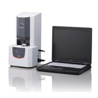
The BioSpec-nano only requires 1 µL (path length: 0.2 mm) or 2 µL (path length: 0.7 mm) of sample, which is pipetted onto the measurement plate. For ultra-small sample volumes no standard rectangular cell is needed, although for applications that are not volume-limited, an optional rectangular cell adapter is available. With the most automatic functionality available on the market, sample mounting, measurement and cleaning are conducted by the instrument. The operators need not perform tedious, repetitive, and inconsistent placement of the fiber optic element and manual cleaning required in other instruments. Sample measurement time is 3 seconds.
Dedicated software allows simple and quick operation of the BioSpec-nano. The start, exchange wiper, list, detail, save CSV/PDF and display PDF operations are displayed on the toolbar according to the user’s procedure and available with the click of a button.
The BioSpec-nano is an ideal instrument for research in genomics, molecular biology, agriculture, medical science and food quality.
Active Questions & AnswersAsk a Question
Error light is on
UV/VIS Spectrophotometers Service ProvidersView All (32)
Documents & ManualsView All Documents
Features of BioSpec-nano
Ultra-small Sample Volumes Perform analysis with 1µL to 2µL samples. For ultra-small sample volumes no standard rectangular cell is needed although for applications that are not volume-limited, an optional rectangular cell adapter is available.
Automated Analysis With “drop-and-click” functionality, analysis has never been easier. Simply drop the sample onto the target and click the button.
Easy Operation/Maintenance Sample mounting, measurement and cleaning are conducted by the instrument, eliminating the need for operators to perform the tedious, repetitive, and inconsistent placement of the fiber optic element and manual cleaning required in other instruments.
Speed Measurement takes seconds, and a series of samples can be analyzed while confirming spectra in the Detail view mode.
Protein Analysis Capability Enables the quantitation of simple proteins and proteins labeled with Cy3 and other labels. Below is an example of the quantification of Lysozyme labeled with the dye AlexaFluor-546. The concentrations of protein, label and the label ratio are easily determined.
General Specifications
| Bandwidth | 3 nm |
| Depth | 214 mm |
| Height | 417 mm |
| Width | 210 mm |
| Power Requirements | AC 100 V / 120 V / 220 V / 230 V / 240 V, 50 / 60 Hz, 40 VA |
| Optical System | Monochromator: Holographic grating |
| Photometric Range | 0 to 1.5 Abs |
| Weight | 7 kg |
| Light Source | Xenon flash lamp |
| Photodetector | Diode Array |
Additional Specifications
Wavelength accuracy: ±1 nm
Pathlength: 0.2 mm, 0.7 mm (manual selection)
Photometric value unit: OD (Optical Density), absorbance converted with 10 mm pathlength
Option cell: Available (pathlength: 5 mm, sample volume: 2 mL) Pathlength lever switched to Option (5 mm) position
Sample volume: 1 µL min. (pathlength: 0.2 mm), 2 µL min. (pathlength: 0.7 mm)
Auto wiping function: Provided
Sample mount function: Auto
Spectrum measuring time: 3 sec
Quantitation range (OD, dsDNA concentration):
Pathlength 0.2 mm, 1 to 75 OD, 50 to 3,700 ng / µL
Pathlength 0.7 mm, 0.3 to 21 OD, 15 to 1,000 ng / µL
Optional 5 mm pathlength cell, 0.04 to 3 OD, 2 to 150 ng / µL