Thermo Scientific - myECL™ Imager
One-touch image capture and analysis for protein and nucleic acid gels and blots.
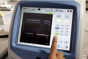
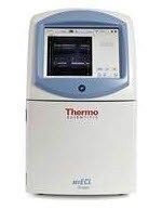
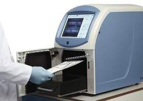
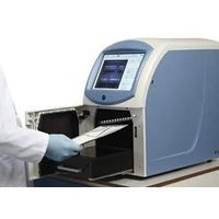
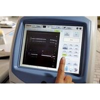
The myECL Imager revolutionizes image acquisition of protein and nucleic acid blots and gels detected via chemiluminescent, colorimetric or UV light-activated fluorescent substrates or stains. The compact benchtop instrument uses UV and visible light transillumination, specialized filters and advanced CCD camera technology to capture images with high sensitivity and dynamic range. The large, 10.4-inch touchscreen and on-board computer provide an elegant user-interface to program acquisition settings and manage (store and share) image files. Also included with the instrument is a five-computer license for the Thermo Scientific myImageAnalysis™ Software, a complete and powerful analysis tool.
Active Questions & AnswersAsk a Question
Recent Questions & Answers
Asked byblksabbath
Need Equipment Support?
Documents & ManualsView All Documents
Features of myECL™ Imager
- One-touch image acquisition – press any one of several optimized presets in each mode and the imager does all the rest; no focusing or camera settings need to be adjusted
- Multi-exposure feature – automatically capture a series of images using up to five different preset or user-defined exposure times
- Automatic visible image capture – system automatically takes a corresponding visible image with every chemiluminescent image exposure; allows overlay alignment with prestained MW markers
- Remote Tech Support access – share your myECL Imager screen in a live session with Technical Support to receive immediate help while using the instrument
- Live camera setting – in any mode see a live view of the illuminated platform on the display screen while the door is open so you can place and center the sample
- Shoot-and-review convenience – imager keeps the last five captured images immediately available in on-screen tabs so you can quickly review, compare, choose and make adjustments to results
- Interactive Chemi – automatically calculates the exposure time of a Western blot with maximum dynamic range and minimal pixel saturation from a short, 15-second exposure image
- File manager – easily copies, deletes, exports and edits image information of one or more image files in multiple gallery folders
- Create dark and bias images – creates new dark and bias master files to compensate for noise coming from the CCD camera during image acquisition
- Adjust image – adjust the black, white and gamma levels of acquired images to increase sample visibility
- Intensity display – select a point of interest on the acquired image to view the pixel intensity and pixel coordinates of the corresponding region
Includes: The myECL Imager ships equipped with 306nm bulbs in the UV transilluminator and the Orange Filter installed in Filter Position 2 (necessary for ethidium bromide and SYPRO™ Orange stains). Also included are the following accessories: Chemiluminescence Exposure Screen, White Light Conversion Screen, UV Exposure Screen, Imaging Reference Target, Touchscreen Stylus with Holder, 2 GB USB Flash Drive, User Manuals and Quick Start Guides. Each imager purchase also includes a five-computer network license for the myImageAnalysis Software.
Recommended for: Chemiluminescent Western blots (ECL substrates); Stained protein gels (coomassie, silver, UV light-excited fluorescent stains); Chemiluminescent nucleic acid blots (e.g., EMSA and Southern blotting using biotin probes, streptavidin-HRP and ECL substrates); Stained nucleic acid gels (ethidium bromide and other UV light-excited fluorescent stains)
General Specifications
| Depth | 56 cm |
| Height | 53 cm |
| Width | 31 cm |
| Weight | 29.5 kg |
Additional Specifications
CCD Camera
16-bit, 4.2 megapixel; thermoelectrically regulated at -25°C (±0.1)
Lens
50mm, f/0.95
Array size (pixels)
2048 x 2048
Pixel size
7.4 x 7.4µm
Field of view
15.0 x 15.0cm
Dynamic range
>4.0 orders of magnitude
Binning modes
1 x 1, 2 x 2, 3 x 3 (default), 4 x 4, 8 x 8
Image capture modes
Chemiluminescence, UV transilluminator, epi-white light
Image exposure modes
Automatic or manual
Image file format
TIFF (16-bit grayscale)
Excitation source
306nm UV transilluminator; Epi-white light
Filter wheel
Motorized, 4 position
Computer
Internal with 250 GB hard drive†
Touchscreen display
10.4-inch LCD
Ports
3 USB, 1 network
†Approximately 200 GB are available for storage of acquired images, providing storage for more than 200,000 image files captured using the default image acquisition setting (3 x 3 binning).

