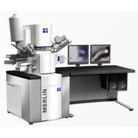
ZEISS - Merlin
Manufactured by ZEISS
Complete image analysis and characterization of biological samples.
Based on the new GEMINI II column and the ZEISS Complete Detection System, MERLIN is the ideal solution for complete image analysis and characterization of biological samples. With the highest beam current in a nanometer size spot, MERLIN provides the fastest imaging of large fields of view at FE-SEM resolution, perfect for streamlined imaging of large samples such as cells or tissue sections of brain, kidney, pathology, plant and forensics.
Image collection speed is further enhanced by sample transfer rates of less than 60 seconds. A simultaneous view of up to four detector signals allows for image comparison without alignment changes for all non-conductive, biological materials due to MERLIN’s unique charge compensation with in-situ cleaning. Novice users are able to achieve professional results using the automated column alignment of MERLIN which quickly attains optimum imaging conditions.
In combination with the ATLAS software, high resolution, extreme field of view imaging is possible with MERLIN, with image sizes of up to 32k by 32k pixels, perfect for examining large sections from brain, kidney, nanopathology and forensic samples or other tissues. MERLIN, in combination with the 3View ultramicrotome, can also be used for high-resolution, 3D-imaging of tissues and even whole organisms, such as Drosophila, zebrafish or plants.
Active Questions & AnswersAsk a Question
There are no current Discussions
Electron Microscopes Service ProvidersView All (8)
Documents & ManualsView All Documents
Features of Merlin
New GEMINI II column and the ZEISS Complete Detection System
- Complete image analysis and characterization of biological samples
- Fastest imaging of large fields of view at FE-SEM resolution
- Streamline imaging of large samples such as cells or tissue sections of brain, kidney, pathology, plant and forensics
Image collection speed with transfer rates of less than 60 seconds
- Image comparison without alignment changes for all non-conductive, biological materials
- Quickly attain optimum imaging conditions
ATLAS Large Area Imaging
- Acquire images up to 32k x 32k pixels with dwell times from 100 nanoseconds to > 100 seconds
- Create large image montages, automatically moving from image tile to tile, resulting in extreme field of view
- Reduces the number of tiles resulting in less beam damage and increased analytical performance
- Examie large sections from brain, kidney, nanopathology and forensic samples or other tissues
General Specifications
| Magnification | 12 to 2000000 x |
| Accelerating voltage | 0.02 to 30 kV |
| Electron Microscope Type | SEM |
| Probe Current | 10 pA up to 300 nA (depending on system configuration) |
| Microscope Type | Electron |
Additional Specifications
Resolution (optimal WD): 0.8 nm @ 15 kV, 1.4 nm @ 1 kV, 3.0 nm @ 20 kV at 10 nA, WD = 8,5mm, & 0.6 nm @ 30 kV (STEM mode)
Electron Emitter: Thermal field emission type, stability > 0,2%/ h